
History:
An eleven-year-old girl with a mass at the left side of the head for many years
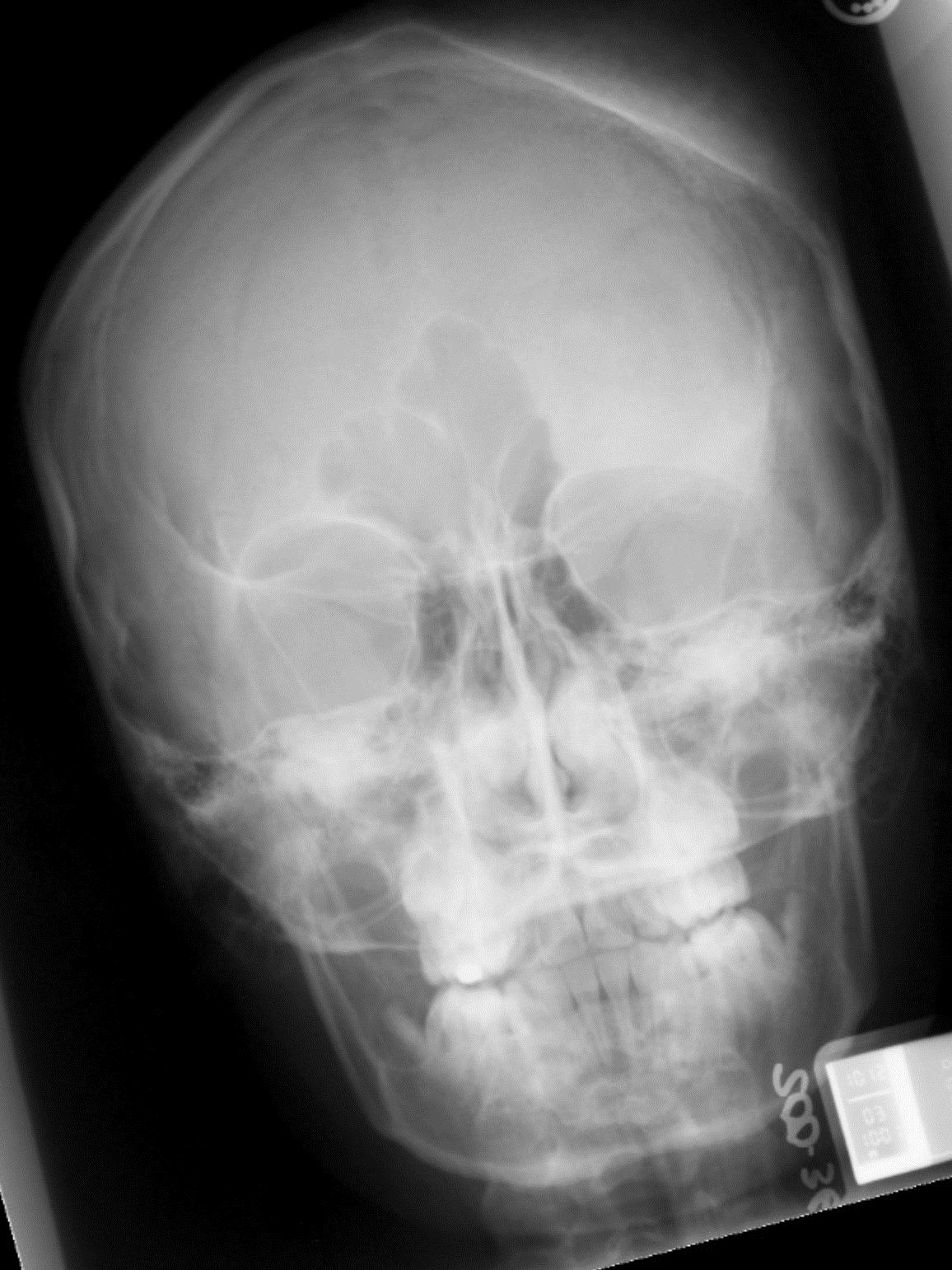
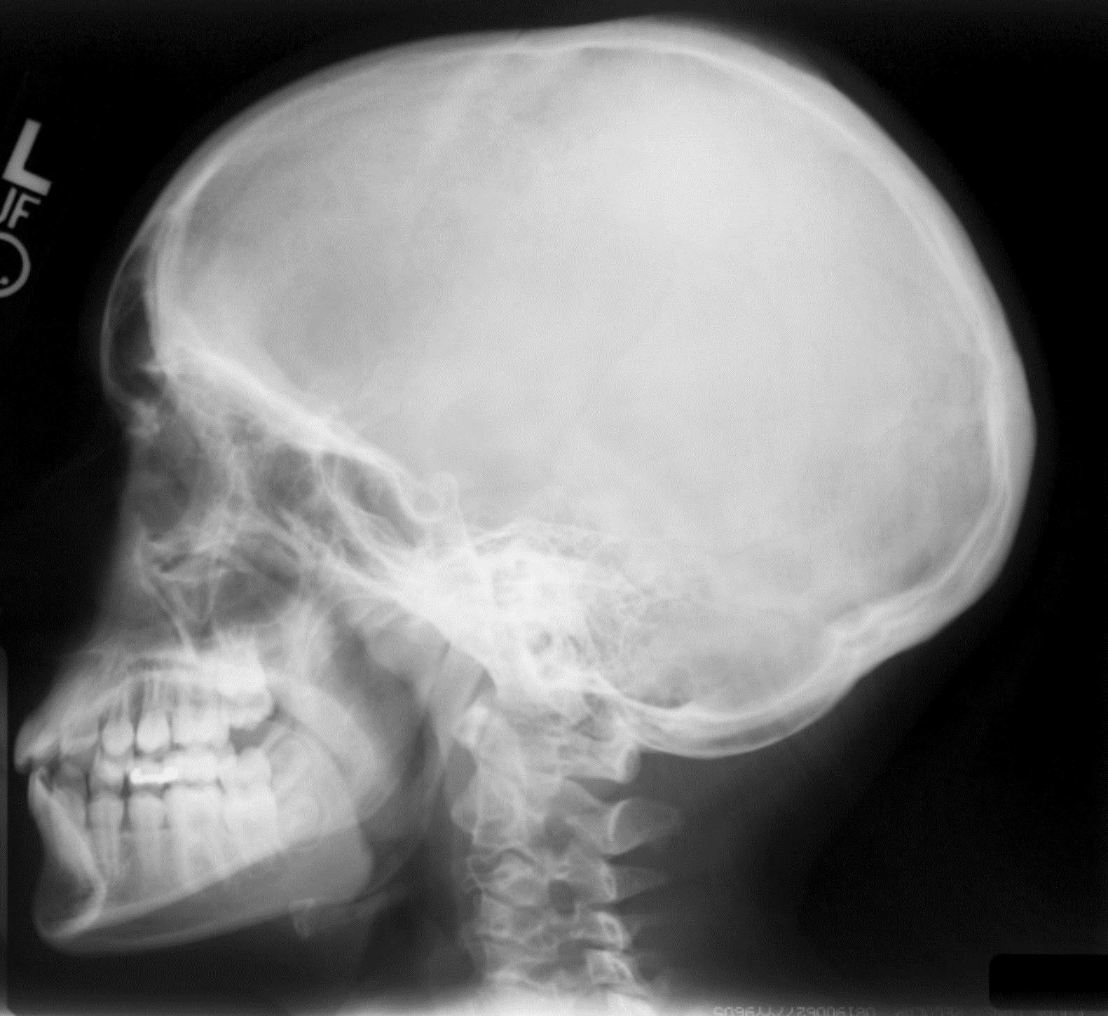

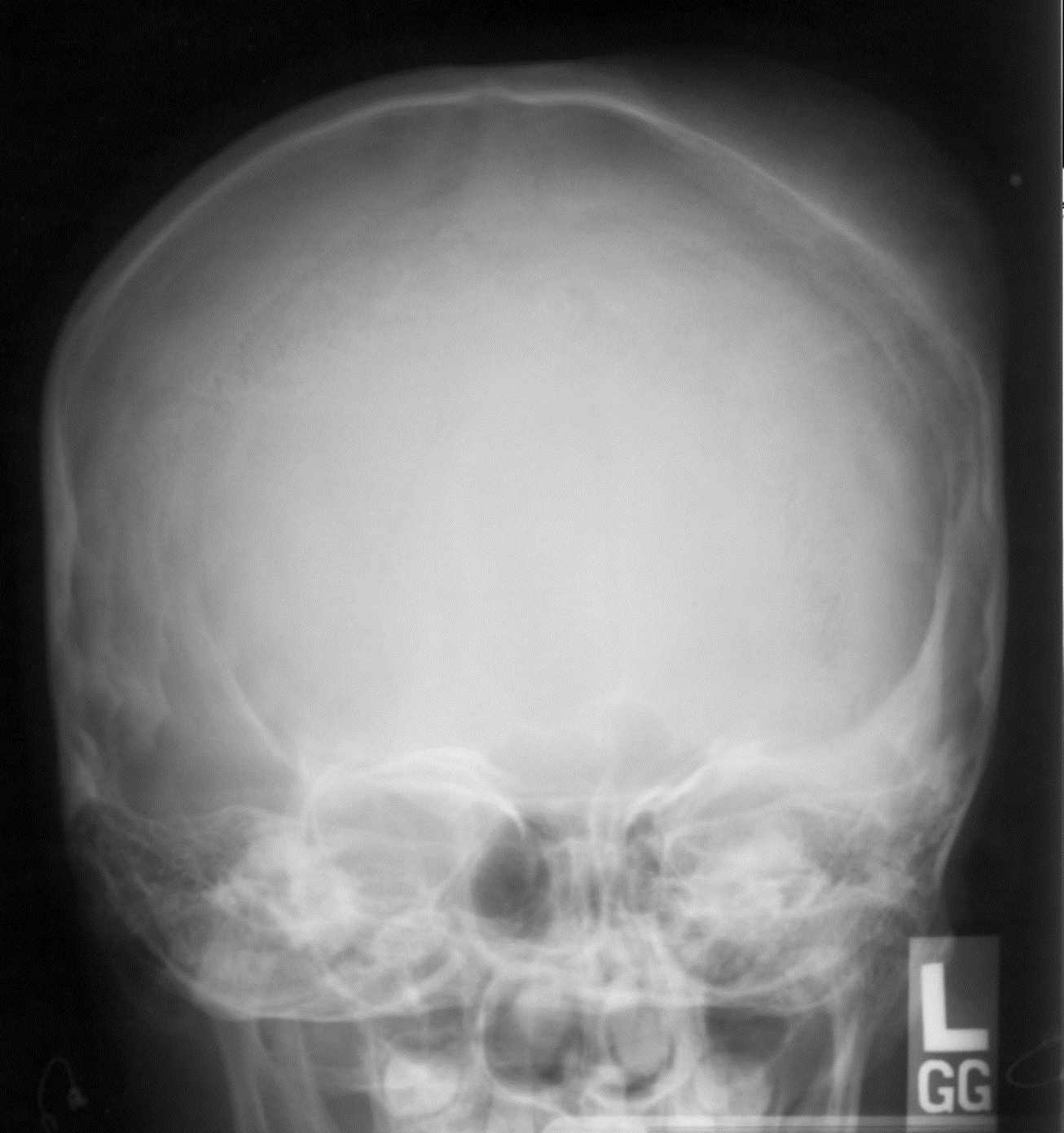
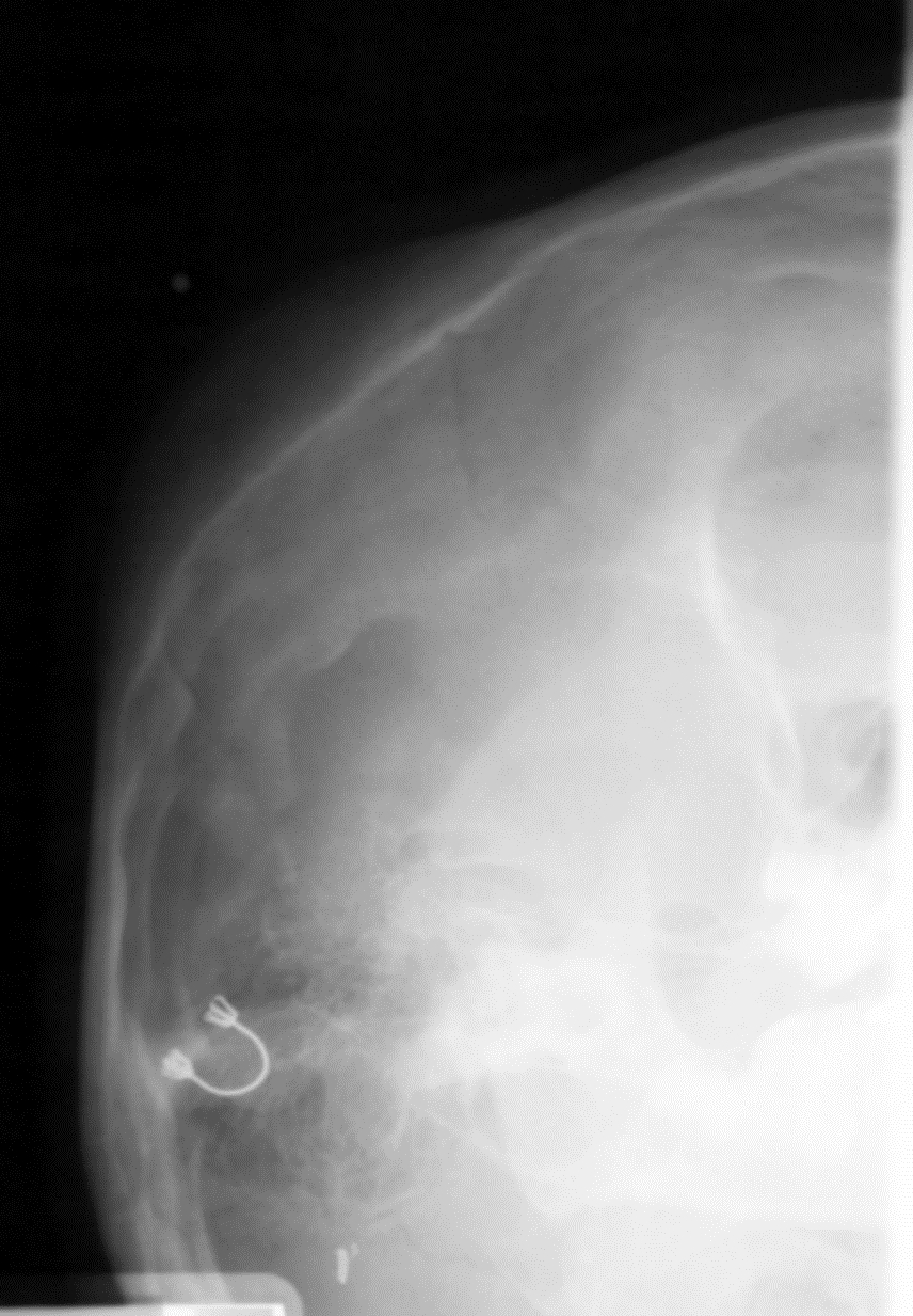

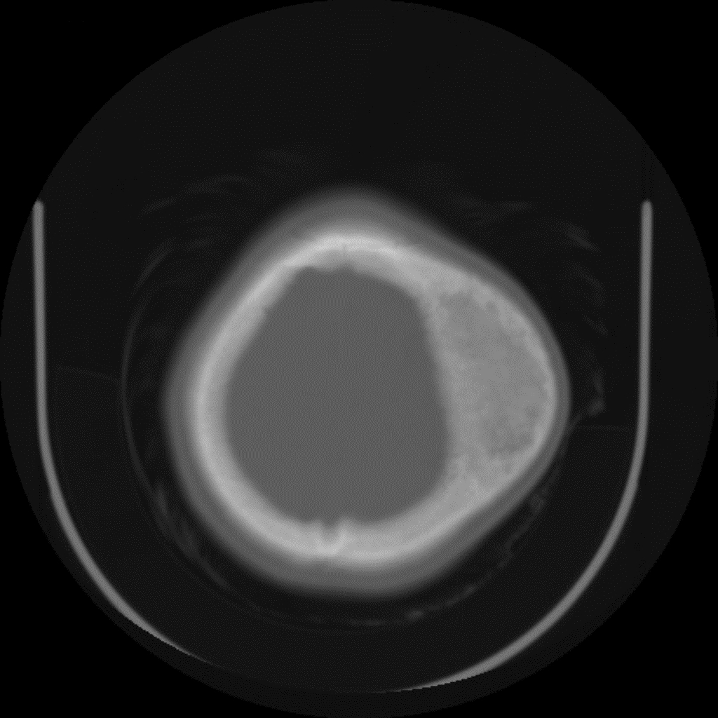
1. What is the most likely diagnosis?
A. Osteosarcoma
B. Paget’s disease
C. Fibrous dysplasia
D. Calvarial metastases
E. Subgaleal hematoma
Show correct answer
2. In relation to facial involvement of fibrous dysplasia, which statement is false?
A. The mandible is more common involved than the maxilla.
B. Involvement of facial bones is more commonly found in the polyostotic form than in the monoostotic form.
C. Cranial nerve palsy is one of the manifestations of fibrous dysplasia.
D. Mucoceles have been reported related to fibrous dysplasia in facial bone.
E. A transformation to osteosarcoma, fibrosarcoma or chondrosarcoma is rare.
Show correct answer
3. In relation to fibrous dysplasia, which statement is false?
A. On T1WI, the signal intensity of fibrous dysplasia is similar to that of the skeletal muscle.
B. On T2WI, the signal intensity of fibrous dysplasia is variable, hyper- or hypointense.
C. Fluid-fluid levels have been described in fibrous dysplasia.
D. Soft tissue extension is common in fibrous dysplasia.
E. Peripheral enhancement may be seen after gadolinium administration.
Show correct answer
Show discussion
References:
1. Robson CD, Kim FM, Barnes PD. Head and neck. In: Kirks DR, Griscom NT. Practical pediatric imaging: diagnostic radiology of infants and children. Philadelphia: Lippincott-Raven Publisher, 1998:225.
2. Dahnert W. Bone and soft tissue disorders. In: Dahnert W. Radiology review manual. Philadelphia: Lippincott Williams and Wilkins, 1996:63.
3. Fletcher BD. Benign and malignant bone tumors. In: Kuhn JP, Slovis TL, Haller JO. Caffey’s pediatric diagnostic imaging. Philadelphia: Mosby, 2003:2387
4. Swischuk LE. Head, brain and meninges. In: Swischuk LE, eds. Imaging of the newborn, infant and young child. Maryland: Willianms and Wilkins, 1997:1007.
5. Yuceer N, Kutluhan A, Bekerecioglu, Arslan H, Akman. Polyostotic fibrous dysplasia with craniofacial localization presenting with frontal lobe compression in a 14-year-old girl. Acta Neurochair 1999;141: 203-7.
6. Sirvanci M, Karaman K, Onat L, Duran C, Ulusoy, OL. Monostotic fibrous dysplasia of the clivus MRI and CT findings. Neuroradiology 2002; 44: 847-50.